

In the human colon, Leb antigen expression evolves through development and cancer progression. While Leb antigens are prevalent in the fetal colon, their presence diminishes in the distal colon postnatally, with limited expression in the proximal colon in adulthood. In contrast, Lea antigens maintain a consistent distribution post-birth. Colorectal carcinoma advances through stages, including hyperproliferation, dysplasia, adenoma formation, carcinoma development, and metastasis. Immunohistochemical studies reveal Leb and Ley antigen resurgence during adenoma stages, intensifying as tumors progress, notably in distal colon cancer. This Leb antigen resurgence is a valuable marker for distinguishing malignancy, aiding early diagnosis and differentiation from non-malignant conditions.
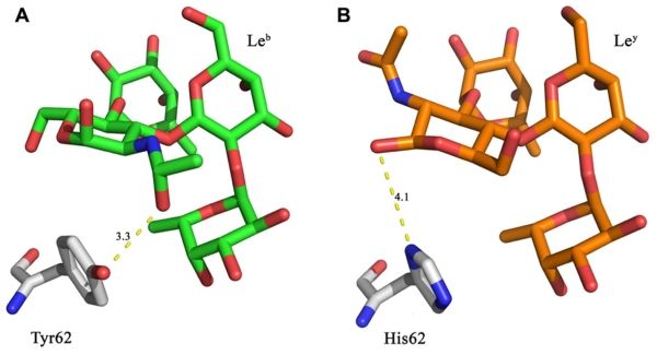
 Fig.1 (A) Leb is within hydrogen bonding distance (3.3 Å) of Tyr62 from the LLYlecwt domain. (B) A Y62H mutation was designed to retain similar structural properties to tyrosine, with the possibility that it may also result in a hydrogen bond to the Ley antigen. (Lawrence, et al., 2012)
According to different glycosidic bonds, Lewis antigens are classified as type I and type II. We have developed the Glyco™ Vaccine Development Service Platform to provide high-quality Tumor-Associated Carbohydrate Vaccine Development services to our clients around the world, specifically the Tumor-Associated Lacto-series Antigen Production Service. It is a common production strategy to regulate the production of Leb antigen products by regulating the gene expression of the relevant enzymes according to the Leb biosynthetic pathway. Here are the pathways on which our services are based.
The H type I structure is generated by adding fucose (Fuc) to the type I structure through the action of α1,2fucosyltransferases (α1,2Fuc-Ts). Subsequently, the Le enzyme adds Fuc to the H type I structure to create Leb antigens. Our previous research has shown that Leb expression on erythrocytes, as well as secretor status determination, is exclusively governed by the Se enzyme. On the other hand, the H enzyme is solely responsible for determining ABH antigen expression in erythrocytes. In colon tissues, both α1,2Fuc-Ts, namely the Se and H enzymes, are expected to participate in the synthesis of H-type I structures, eventually leading to the production of Leb antigens.
 Fig.2 Lewis antigens biosynthetic pathways. (Ma, et al., 2021)
Here is the standard process of our service, the Leb antigens are formulated into a vaccine. Depending on the type of vaccine, they may be combined with adjuvants, carriers, or other immune-stimulating molecules to enhance their immunogenicity. Leb antigen-based vaccines hold promise in cancer immunotherapy, offering a targeted approach to combat specific cancers that overexpress these antigens. They are part of ongoing efforts to harness the immune system's capabilities to fight cancer and improve patient outcomes. We look forward to your contacting us to inquire about the details!
 Fig.3 A series of enzymatic reactions that result in the synthesis of Leb antigens. (CD BioGlyco)
Technology: Tumor Marker, Glycosyltransferase
Journal: Cancers
IF: 5.2
Published: 2020
Results: CA19.9, often used as a tumor marker, was believed to be significantly elevated in gastrointestinal cancer due to abnormal glycosylation. New findings, however, indicate that the detection of CA19.9 aligns with biochemical and molecular data. Lea and Leb, related carbohydrate antigens, are absent in colon cancer because of suppressed galactosyltransferase activity. In pancreatic cancer, no marked difference in CA19.9 or relevant glycosyltransferases exists. Lewis antigens are expressed in a pattern influenced by glycosyltransferase levels, determined genetically and epigenetically. Elevations in circulating antigens result from duct obstruction and polarity loss in malignant cells. These antigens show promise for monitoring pancreatic cancer patients negative for CA19.9, but not as a CA19.9 alternative. Personalized patient studies hold potential for translational research and clinical applications.
 Fig.4 Structure of type 1 chain Lewis antigens and biosynthesis in gastrointestinal tissues. (Indellicato, et al., 2020)
CD BioGlyco is a company with extensive glycan chemistry research experience and aims to be clients’ best glycan research cooperator. Please do not hesitate to contact us if you are interested in our services and would like to have a further discussion with us.
References
